Stapled peptides are peptides chemically constrained into stable alpha-helical conformations through covalent side-chain crosslinks, a technique that directly addresses the three core failures of therapeutic peptides: protease degradation, poor membrane permeability, and loss of bioactive conformation. The standard approach uses non-natural amino acids bearing olefin side chains, crosslinked by ruthenium-catalyzed ring-closing metathesis (RCM) using the Grubbs second-generation catalyst. Staple positions follow defined spacing rules, with i, i+4 spanning one helical turn and i, i+7 spanning two. Understanding stapled peptide design explained at this mechanistic level is the prerequisite for making rational decisions in therapeutic peptide development.
What is stapled peptide design and how does it work chemically?
Stapled peptide synthesis begins with the incorporation of non-natural alpha-methyl amino acids bearing terminal olefin side chains at two defined sequence positions. These positions, typically i, i+4 or i, i+7, are selected to place both reactive residues on the same face of the nascent helix. The Grubbs second-generation ruthenium catalyst then performs ring-closing olefin metathesis, forming a covalent all-hydrocarbon bridge that locks the backbone geometry.
The i, i+4 staple spans a single helical turn and suits shorter peptides or sequences where one face needs reinforcement. The i, i+7 configuration spans two turns, providing greater rigidity and a larger hydrophobic surface. Choosing between them depends on the target interface geometry and the length of the peptide segment you need to stabilize. For sequences requiring exceptional rigidity, double stapling or "stitched" peptides incorporate two consecutive crosslinks, though this significantly increases synthetic complexity.

After RCM, the double bond in the hydrocarbon staple can be optionally hydrogenated to improve metabolic stability and reduce potential reactivity. This step is often overlooked in early-stage work but becomes relevant when advancing candidates toward in vivo studies. Strict anaerobic conditions are required throughout RCM because ruthenium catalysts are oxygen-sensitive, which adds manufacturing overhead and contributes to the cost of scaling this chemistry beyond research quantities.
Pro Tip: When designing your staple position, map the helical wheel projection of your target-binding peptide first. Place the staple exclusively on the solvent-exposed face, opposite the residues that contact the target. Incorrect placement abolishes binding even when helicity increases.
The synthesis workflow follows a numbered logic:
- Select the target-binding sequence from structural data (X-ray, cryo-EM, or NMR).
- Identify the binding face and mark residues for staple placement on the opposing face.
- Incorporate alpha-methyl, alpha-alkenyl amino acids at the chosen i, i+4 or i, i+7 positions using standard Fmoc solid-phase peptide synthesis.
- Perform on-resin or in-solution RCM with Grubbs second-generation catalyst under inert atmosphere.
- Cleave, purify by RP-HPLC, and confirm staple formation by mass spectrometry and circular dichroism.
What are the biological benefits of stapled peptides in drug discovery?
Stapling produces measurable, reproducible improvements across the three pharmacological parameters that most often disqualify peptide drug candidates. Alpha-helical content increases from below 20% in linear peptides to 60 to 90% in stapled forms. That structural pre-organization is the origin of most downstream benefits.
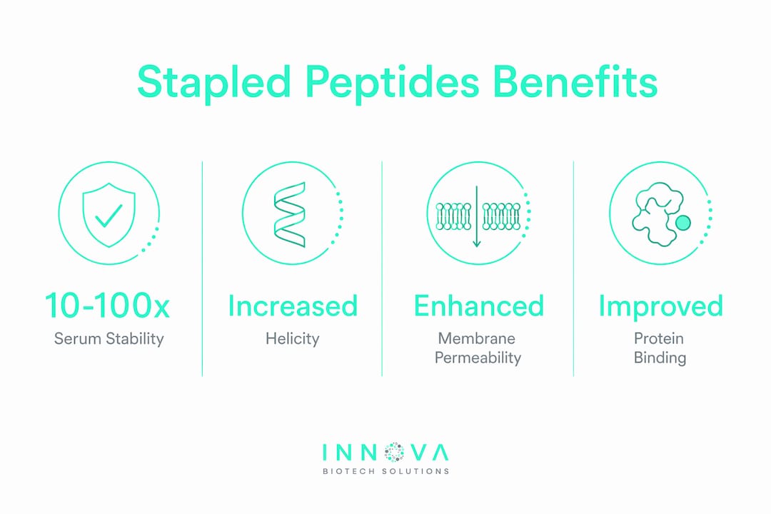
Protease resistance is the most immediately practical gain. Hydrocarbon staples physically block protease access to backbone cleavage sites, and the alpha-methyl substitution at staple positions further resists enzymatic attack. The result is 10- to 100-fold improvements in serum half-life compared to linear analogs. For drug developers, this translates directly into viable dosing intervals and systemic exposure levels that support in vivo efficacy studies.
Binding affinity improvements are equally significant. By pre-organizing the peptide into its bioactive conformation, stapling eliminates the entropic penalty of folding upon target engagement. Hydrocarbon staples increase binding affinity by 5- to 5000-fold depending on the target and sequence context. This range reflects how much the linear peptide was paying in conformational entropy before stapling.
Stapled peptides overcome three simultaneous pharmacological barriers: protease degradation, poor membrane permeability, and loss of bioactive conformation. No other single modification achieves all three at once.
The membrane permeability benefit is mechanistically distinct. The hydrophobic staple surface creates an amphipathic helix geometry that facilitates direct membrane translocation, enabling access to intracellular protein-protein interactions. There are over 650,000 intracellular protein-protein interactions in the human proteome, the vast majority of which remain inaccessible to conventional small molecules. Stapled peptides represent one of the few credible strategies for targeting this space.
The clinical candidate ALRN-6924, a stapled peptide inhibitor of the p53-MDM2 and p53-MDMX interactions developed by Aileron Therapeutics, demonstrates what this technology can achieve in practice. It has progressed through Phase II trials in hematologic malignancies, providing direct evidence that stapled peptides can survive the translational gauntlet from bench to clinic. Beyond oncology, stapled peptides have expanded into metabolic disease, infectious disease, and wound healing, reflecting the generality of the approach.
You can find a detailed breakdown of how peptide secondary structure influences these pharmacological outcomes in Innovabiotech's published resources.
How do computational tools and experimental assays work together in stapled peptide optimization?
Computational design accelerates stapled peptide development by narrowing the sequence and staple-position space before synthesis begins. Molecular dynamics simulations model helical propensity and predict how different staple configurations affect backbone geometry and target engagement. Free energy perturbation calculations estimate relative binding affinities across staple variants, guiding prioritization of candidates for synthesis.
The practical limitation is significant: molecular dynamics simulations frequently misestimate stapled peptide helicity and binding affinity, requiring iterative empirical validation to refine designs. Force field parameters for non-natural alpha-methyl amino acids are less mature than those for canonical residues, and the conformational sampling of constrained peptides presents specific challenges. Treat computational outputs as hypothesis generators, not as final answers.
Experimental validation requires a defined assay cascade:
- Circular dichroism (CD): Quantifies helical content in aqueous buffer and in membrane-mimicking environments. This is the first filter after synthesis.
- Serum stability assays: Measure half-life in human or mouse serum. Protocols for peptide stability testing are well-established and should be run in parallel with binding assays.
- Fluorescence polarization or SPR: Quantifies binding affinity to the target protein with defined Kd values.
- Cell permeability assays: Quantitative cytosolic delivery assays using split-GFP or chloroalkane penetration assays are now preferred over qualitative confocal imaging, which cannot distinguish endosomal trapping from true cytosolic delivery.
- Cell viability panels: Screen for cytotoxicity across a panel of cell lines early, before investing in target-specific cellular assays.
Pro Tip: Run CD in both phosphate buffer and 50% trifluoroethanol. The TFE condition reveals maximum helical potential, while buffer-only CD reflects the actual solution conformation your peptide adopts under physiological conditions. The gap between the two tells you how much conformational entropy remains after stapling.
Structural biology data from X-ray crystallography or cryo-EM of the target interface is the highest-value input for computational design. Innovabiotech's computational modeling services integrate structural data directly into staple placement workflows, reducing the number of synthesis-test cycles needed to reach an optimized candidate.
What linker engineering strategies improve membrane permeability and safety?
The all-hydrocarbon staple is the established standard, but it is not the only option. Researchers now deploy a range of alternative linker chemistries to tune the physicochemical profile of stapled peptides for specific delivery challenges.
| Linker type | Key property | Primary trade-off |
|---|---|---|
| All-hydrocarbon (olefin) | High helicity, protease resistance | Lipophilicity can cause aggregation |
| Aryl-thioether | Improved aqueous solubility | Reduced helical stabilization vs. hydrocarbon |
| Charged/polar linkers | Enhanced solubility, reduced toxicity | Lower membrane permeability |
| Stimuli-responsive (redox, pH) | Controlled release at target site | Increased synthetic complexity |
| Lactam bridges | Aqueous compatibility | Less hydrophobic surface for membrane crossing |
The central tension in linker engineering is the relationship between lipophilicity and cellular uptake. Increasing hydrophobicity improves membrane translocation up to a threshold, beyond which high lipophilicity causes aggregation, serum protein binding, and reduced cellular potency. This solubility cliff is one of the most common failure modes in stapled peptide optimization.
Stimuli-responsive reversible stapling addresses a specific problem: the same hydrophobic surface that drives membrane crossing can also cause off-target interactions once the peptide is inside the cell. Reversible stapling separates delivery enhancement from target binding, allowing the staple to open under intracellular reducing conditions and releasing a more polar, target-selective peptide. This approach is still early-stage but represents a mechanistically sound solution to the permeability-selectivity trade-off.
"Chameleonic" linker designs exploit the ability of certain chemical motifs to adopt different conformations depending on their environment, presenting a hydrophobic face in the membrane and a more polar face in aqueous solution. High-throughput screening platforms including mRNA display, phage display, and DNA-encoded chemical libraries are increasingly used to identify sequences that combine intrinsic helicity with chameleonic behavior, reducing the reliance on purely rational design.
Pro Tip: When evaluating membrane permeability, use quantitative cytosolic delivery assays rather than confocal imaging. Fluorescence in the cell does not confirm cytosolic access. Endosomal trapping produces the same visual result and leads to false confidence in permeability data.
Intramolecular hydrogen bond shielding (IMHB) is another post-screening refinement strategy. Methylating backbone amide NH groups reduces the desolvation penalty for membrane crossing without adding bulk or changing the staple chemistry. Combining IMHB optimization with a well-placed hydrocarbon staple can push permeability into ranges competitive with small molecules for certain target classes.
Key takeaways
Stapled peptide design succeeds when chemical stapling, staple placement, and physicochemical optimization are treated as a single integrated problem rather than sequential steps.
| Point | Details |
|---|---|
| Staple position is critical | Place crosslinks on the solvent-exposed face only; wrong placement abolishes binding regardless of helicity gains. |
| Biological gains are quantifiable | Stapling raises helical content to 60 to 90% and extends serum half-life by 10- to 100-fold versus linear peptides. |
| Computation guides but does not replace experiment | Molecular dynamics predictions require validation by CD, serum stability, and quantitative cytosolic delivery assays. |
| Linker chemistry controls the permeability-toxicity balance | Hydrocarbon staples maximize helicity; stimuli-responsive and charged linkers address solubility and off-target risks. |
| Clinical translation remains challenging | Scaling RCM under anaerobic conditions is costly, and immunogenicity assessment must be built into the development plan. |
Why staple placement still trips up experienced teams
After working through stapled peptide design projects across oncology and metabolic disease targets, the pattern I see most often is not a chemistry failure. It is a design failure that happens before synthesis begins. Teams optimize helical content and binding affinity in isolation, then discover that the staple they placed for maximum rigidity sits directly on a residue that contacts the target. The peptide is beautifully helical and completely inactive.
The computational tools available today, including molecular dynamics and free energy calculations, are genuinely useful for narrowing the design space. But prediction inconsistencies between in silico models and experimental outcomes are not edge cases. They are routine. The force field limitations for non-natural amino acids mean you should always treat a computational prediction as a ranked shortlist, not a final answer.
The other issue I see underestimated is the cytosolic delivery question. Qualitative confocal imaging is still used in too many programs as the primary evidence for cell permeability. Endosomal trapping looks identical to cytosolic delivery under a fluorescence microscope. Programs have advanced candidates based on this data and then failed to show target engagement in cellular assays. Quantitative cytosolic delivery assays should be standard, not optional.
The most promising direction I see is stimuli-responsive stapling combined with quantitative delivery profiling from the earliest stages. It separates the delivery problem from the binding problem in a way that hydrocarbon staples alone cannot. Foldamer-inspired designs that go beyond simple alpha-helices are also worth watching, particularly for targets where the binding interface is not a clean helix-mediated interaction. The field is moving fast, and the teams that integrate structural biology, quantitative cell biology, and computational design from day one are the ones producing candidates that survive into the clinic.
— Hooman
How Innovabiotech supports your stapled peptide program
Innovabiotech's peptide design and optimization services are built specifically for research teams working on therapeutic peptide candidates that require non-natural amino acid incorporation, advanced staple configuration, and bioinformatics-guided validation. The team integrates structural biology data, molecular dynamics modeling, and empirical assay design into a single workflow, reducing the number of synthesis cycles needed to reach a validated lead.

Whether you are positioning a first staple on a new target or optimizing a candidate series for cytosolic delivery and serum stability, Innovabiotech provides the computational and experimental expertise to move faster with higher confidence. Every project is scoped to your specific target class, sequence constraints, and development timeline. Contact Innovabiotech to discuss how a tailored stapled peptide design program can accelerate your therapeutic candidate pipeline.
FAQ
What is a stapled peptide?
A stapled peptide is a synthetic peptide chemically constrained into an alpha-helical conformation by a covalent crosslink, typically an all-hydrocarbon bridge formed by ring-closing metathesis between two non-natural olefin-bearing amino acids. This structural lock improves protease resistance, membrane permeability, and target binding affinity simultaneously.
What are the i, i+4 and i, i+7 staple positions?
The i, i+4 position places the two crosslinked residues one helical turn apart, while i, i+7 spans two turns. The choice depends on the length of the helical segment being stabilized and the geometry of the target interface. Both positions require the crosslinked residues to sit on the same face of the helix.
How much does stapling improve serum stability?
Stapled peptides show 10- to 100-fold improvements in serum half-life compared to their linear counterparts. The hydrocarbon bridge physically blocks protease access to backbone cleavage sites, and the alpha-methyl substitution at staple positions adds further resistance to enzymatic degradation.
Why do computational predictions for stapled peptides require experimental validation?
Molecular dynamics force fields for non-natural alpha-methyl amino acids are less accurate than those for canonical residues, causing frequent misestimation of helicity and binding affinity. Circular dichroism, serum stability assays, and quantitative cytosolic delivery measurements are required to confirm and refine computationally derived designs.
What is the main challenge in scaling stapled peptide synthesis?
Ring-closing metathesis requires ruthenium catalysts that demand strict anaerobic conditions throughout the reaction, making large-scale manufacturing costly and technically demanding. Immunogenicity assessment of the non-natural amino acid components also adds regulatory complexity during clinical development.
Recommended
- Innova Biotech Solutions: Expert Peptide Design and Optimization Services.bioinformatics validation, custom peptides, peptide design services
- Peptide Secondary Structure: What Researchers Need to Know
- Examples of De Novo Protein Design: 2026 Research Guide
- Peptide Binding Affinity Prediction: A Practical Guide