
The principal types of preclinical screening assays are biochemical (cell-free), cell-based, and in vivo models. The most defensible drug-discovery cascades run them in a tiered sequence: primary screen → orthogonal confirmation → secondary mechanistic assays, with ADME/Tox checks embedded during hit-to-lead rather than saved for later.
Each class has a defined role:
- Biochemical assays — high-throughput hit finding against isolated molecular targets (enzymes, receptors, protein-protein interactions)
- Cell-based assays — pathway-level validation, cytotoxicity, and phenotypic discovery with integrated cellular context
- In vivo models — systemic PK/PD, toxicity, and translational efficacy when no in vitro surrogate suffices
Innovabiotech maps computational screening outputs directly to wet-assay selection, accelerating progression through each cascade stage.
Table of Contents
- What does preclinical screening actually mean for assay selection?
- How do biochemical assays work in high-throughput screening?
- Which cell-based assay format fits your program stage?
- When do you actually need in vivo models?
- How do you design a practical screening cascade?
- How do you pick assays when virtual screening drives the program?
- What QC metrics must you lock before running a screen?
- How does the compute-to-assay iteration loop actually work?
- Innovabiotech supports preclinical screening and computational validation
- Key Takeaways
- The assay selection decision most teams get wrong
- Authoritative sources and further reading
- Innovabiotech: computational screening and assay strategy for biopharma teams
- FAQ
What does preclinical screening actually mean for assay selection?
Preclinical screening is the structured process of evaluating compound activity, selectivity, and safety before any human exposure. The FDA defines two main types of preclinical research: in vitro studies (cell cultures, biochemical systems) and in vivo studies (living organisms), with toxicity assessment required before human testing.
"The choice of either biochemical or cell-based assay design and the particular assay format is a balancing act between two broad areas: ensuring the measured signal provides relevant data to the desired biological process, and supporting reagents that yield robust data in microtiter plate formats where very large compound libraries are screened." — NCBI Assay Guidance Manual
That tension between physiological relevance and throughput is the central decision every R&D team faces. For hit identification, throughput wins. For lead optimization, biological context wins. The practical decision criteria are: What is the biological question? What program stage are you in? How many compounds need testing? And critically, are you validating computational predictions from docking or machine learning?
How do biochemical assays work in high-throughput screening?
Biochemical assays are cell-free systems measuring direct molecular interactions: enzyme activity, receptor binding, displacement, and protein-protein interactions. Detection relies on fluorescence, luminescence, or absorbance readouts, and formats scale readily to 384-well and 1536-well plates.
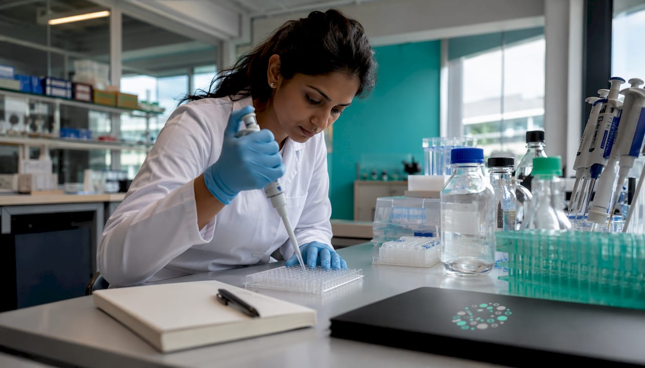
Their throughput advantage is substantial. Because there are no cells to maintain or transfect, biochemical assays are the standard choice for primary HTS of libraries reaching 10⁵–10⁶ compounds. Biophysical readouts like surface plasmon resonance (SPR) and biolayer interferometry (BLI) add binding-kinetics data for tighter hit triage.
The limitation is context. A compound that inhibits a purified enzyme may fail completely in a cellular environment due to membrane permeability, off-target binding, or metabolic conversion. Orthogonal assays using a different detection mechanism on the same target are the standard mitigation, filtering out fluorescent compounds and aggregators before they consume cell-based capacity.
For teams running virtual screening, biochemical binding or displacement assays are the natural first wet-lab step for docking hits. The readout directly mirrors what the docking score predicts.
Pro Tip: Run a counterscreen panel (e.g., a promiscuous-inhibitor filter using a structurally unrelated enzyme) in parallel with your primary biochemical screen. Catching aggregators and redox-active compounds at this stage costs far less than discovering them in cell-based follow-up.
Which cell-based assay format fits your program stage?
Cell-based assays capture integrated cellular responses that biochemical systems cannot: pathway activation, receptor internalization, cytotoxicity, and emergent phenotypes. Cell-based models account for a substantial share of HTS efforts and are indispensable for toxicity and pathway-level assessment.
| Format | Throughput | Biological relevance | Typical readout | When to use |
|---|---|---|---|---|
| Viability/cytotoxicity | High | Moderate | Cell count, ATP, LDH | Early safety triage, selectivity windows |
| Reporter gene (luciferase, GFP) | High | Moderate-high | Luminescence, fluorescence | Pathway modulation, hit validation |
| GPCR/ion-channel functional | Moderate | High | Calcium flux, cAMP, FLIPR | Target-class-specific hit confirmation |
| High-content imaging (HCI) | Moderate | High | Multiparametric morphology | Phenotypic discovery, ML training data |
| 3D spheroids/organoids | Low-moderate | Very high | Viability, histology, omics | Lead optimization, toxicity modeling |
A few practical notes on format selection:
- Reporter assays are the go-to for confirming pathway modulation after a biochemical primary screen; they add cellular context without sacrificing much throughput.
- High-content imaging (HCI) combines automated microscopy with quantitative multiparametric analysis, making it valuable for phenotypic discovery and for generating the rich feature datasets that retrain ML models.
- 3D models (spheroids, organoids, organs-on-a-chip) better mimic human tissue architecture. The FDA Modernization Act 2.0 explicitly authorized these as alternative preclinical techniques, though further validation standards are still being established.
- Plate format matters: 96-well for complex assays, 384-well for standard HTS, 1536-well for ultra-HTS biochemical or simple cell-based formats.
When do you actually need in vivo models?
In vivo studies provide what no cell culture can: systemic PK/PD, organ-level toxicity, and the full metabolic context of a living organism. The FDA requires toxicity assessment in living systems before human trials, and no in vitro surrogate currently replaces that regulatory requirement for most programs.
Rodent models (mouse, rat) are the standard starting point for efficacy and safety studies. Larger species are added when metabolic differences or organ-specific toxicities require it. The FDA Modernization Act 2.0 creates a regulatory path for organoids and organs-on-a-chip as alternatives, but most IND-enabling packages still require traditional animal data.
Statistical design is where most in vivo studies fail. Robust preclinical study design requires power calculations, randomization, allocation concealment, and blinding to produce translational evidence that holds up. Underpowered studies generate noise, not decisions.
Statistical planning target: power ≥ 0.8 and alpha = 0.05, with a predefined primary outcome measure and a calculated minimum significant difference (MSD) locked before data collection starts.
Key design requirements:
- Predefine the primary outcome measure before any dosing begins
- Calculate sample size from MSD and desired power
- Randomize animal allocation and blind outcome assessors
- Run positive and negative controls on every plate or cohort
How do you design a practical screening cascade?
A tiered cascade is the standard framework: primary assays maximize sensitivity and throughput; orthogonal assays remove artifacts using a different detection mechanism; secondary assays establish mechanism, potency, and structure-activity relationships (SAR); lead optimization adds ADME/Tox profiling.
Primary → Orthogonal → Secondary → ADME/Tox → Lead Optimization
Each stage has clear acceptance criteria:
- Primary: Hit rate target, Z' factor ≥ 0.5, defined activity threshold (e.g., >50% inhibition at fixed concentration)
- Orthogonal: Confirmation rate, artifact filter pass rate, counterscreen clearance
- Secondary: IC₅₀/EC₅₀ range, selectivity ratio, mechanistic readout
- ADME/Tox: Caco-2 permeability, cytochrome P450 inhibition, plasma stability, hepatotoxicity flag
Embedding ADME/Tox early during hit-to-lead is one of the highest-leverage decisions a team can make. Poor PK and toxicity are among the most common reasons programs terminate late, and rapid in vitro screens like Caco-2 permeability and CYP inhibition profiling cost a fraction of what a failed in vivo study does.
How do you pick assays when virtual screening drives the program?
When computational outputs are the starting point, assay selection follows a specific logic. The readout must match what the model predicted.
Checklist for computationally driven programs:
- Docking hits → biochemical binding or displacement assay first; confirm the predicted binding mode before investing in cell-based work
- ML-scored compounds → match assay to the phenotype or activity the model was trained on
- Designed proteins/peptides → biophysical binding assays (SPR, BLI, thermal shift) confirm folding and affinity; functional cell assays confirm activity in context
- Enzyme-engineering outputs → enzymatic activity assay with substrate panel; stability assays (thermal denaturation, protease resistance) before cellular testing
Practical constraints to address upfront:
- Throughput alignment: if virtual screening delivers 500 prioritized compounds, a 384-well biochemical assay handles that in a single run; 96-well cell-based formats need batching
- Sample requirements: designed peptides and proteins often arrive in limited quantities; prioritize assays with low material demands (SPR, fluorescence polarization) before committing to cell-based panels
- Iterative speed: the faster the biochemical readout returns, the faster the computational model can be retrained on real activity data
Innovabiotech's virtual screening workflows are designed to deliver prioritized hit lists with the metadata needed to select the right first wet assay directly from the computational output.
What QC metrics must you lock before running a screen?
Assay quality is defined before the screen runs, not after. The key metrics:
- Z' factor: ≥ 0.5 is the accepted threshold for a screenable assay; ≥ 0.6 is preferred for HTS. Z' < 0.5 means the assay needs optimization before scaling.
- Signal window (SW): the separation between positive and negative controls; SW ≥ 2 is a standard acceptance criterion.
- Coefficient of variation (CV): ≤ 20% for intra-plate controls.
- Minimum significant difference (MSD): calculated from replicate runs; the NCBI Assay Guidance Manual recommends MSD < 20% for single-dose screens.
Design rules that matter in practice:
- Predefine the primary outcome measure and power calculation (power ≥ 0.8, alpha = 0.05) before in vivo or critical cell-based studies
- Randomize plate layout to distribute systematic errors; blind readout assessment where possible
- Include both positive and negative controls on every plate, not just at the start of a run
- Distinguish technical replicates (same sample, same run) from biological replicates (independent preparations); both are needed for a defensible result
Pro Tip: Run a pilot plate of 32–48 wells with your positive and negative controls before committing to full HTS. If Z' is below 0.5 on the pilot, fix the assay. Scaling a broken assay generates expensive noise.
How does the compute-to-assay iteration loop actually work?
The recommended workflow is a repeating cycle: compute → prioritize → test → retrain. Each pass through the loop narrows the chemical or protein space and improves model accuracy.
- Compute: generate docking poses, ML scores, or protein design candidates
- Prioritize: rank by predicted affinity, selectivity, or ADME flags; select a testable subset
- Test: run the matched wet assay (biochemical binding for docking hits; HCI for phenotypic ML models; biophysical for designed proteins)
- Retrain: feed confirmed actives, inactives, and assay metadata back into the scoring function or ML model
Specific mappings that work in practice:
- Docking poses → biochemical binding assay → confirmed binders feed SAR model
- ML phenotype predictions → high-content imaging panel → multiparametric readouts retrain the classifier
- Protein engineering cycle → design → SPR/thermal shift → cell functional assay → redesign with activity data
- Bayesian active learning for combination screens → iterative dose-matrix testing → model update
Data integration requirements: standardized result formats, complete plate maps, control values, and compound metadata must accompany every assay batch returned to the computational team. Without that metadata, retraining produces a worse model, not a better one.
Innovabiotech supports preclinical screening and computational validation
Pairing computational triage with the right wet-assay strategy is where most programs either accelerate or stall. Innovabiotech offers the computational layer that makes assay selection precise from the first experiment.
Core services relevant to preclinical screening programs:
- Virtual screening and hit-to-lead optimization — structure-based and ligand-based docking, ML scoring, and prioritized hit lists with assay-ready metadata
- Protein engineering and peptide design — de novo design, stability optimization, and chimeric protein modeling with biophysical validation guidance
- Enzyme optimization — activity and stability profiling workflows for enzyme-target programs
- ADME/Tox advisory — early-stage screening strategy to embed permeability and CYP profiling during hit-to-lead
Every project starts with a technical scoping call to map your target class, library size, and computational outputs to the right cascade. Contact Innovabiotech to scope your screening strategy.
Key Takeaways
A tiered preclinical screening cascade integrating biochemical, cell-based, and in vivo assays with early ADME/Tox and locked QC metrics gives R&D teams the most defensible path from computational hit to lead candidate.
| Point | Details |
|---|---|
| Match assay to question | Biochemical assays for molecular hit finding; cell-based for pathway and toxicity; in vivo for systemic PK/PD. |
| Embed ADME/Tox early | Run Caco-2 permeability and CYP inhibition during hit-to-lead, not after in vivo studies. |
| Lock QC before scaling | Z' factor ≥ 0.5, CV ≤ 20%, and MSD < 20% must be confirmed on a pilot plate before HTS. |
| Statistical rigor is non-negotiable | Power ≥ 0.8 and alpha = 0.05 with a predefined primary outcome measure, per FDA guidance. |
| Innovabiotech accelerates iteration | Virtual screening, protein engineering, and hit-to-lead services map computational outputs to wet-assay selection from day one. |
The assay selection decision most teams get wrong
The conventional wisdom says: run your biochemical primary screen, confirm hits in cells, then worry about ADME. That sequence made sense when computational tools were slow and compound libraries were small. It does not make sense now.
When virtual screening delivers a prioritized list of 200–500 compounds with predicted binding modes, the biochemical primary screen is no longer the bottleneck. The bottleneck is the decision about which cell-based assay to run next, and whether the designed protein or peptide you are testing actually folds and binds before you commit it to a cellular system.
The teams that move fastest treat the compute-to-assay loop as a single integrated workflow, not two separate departments. Assay metadata flows back to the model. The model improves. The next round of wet experiments is smaller and better targeted. That cycle, run tightly, is what separates a 12-month hit-to-lead from a 24-month one.
The other underestimated decision is statistical design for in vivo work. A study powered at 0.6 instead of 0.8 does not just produce a weaker result. It produces a result you cannot trust, which means you run the study again. The cost of that repeat dwarfs the cost of a proper power calculation done before the first animal is dosed.
Authoritative sources and further reading
- FDA Step 2: Preclinical Research — FDA definition of in vitro vs in vivo preclinical studies and regulatory requirements before human testing.
- NCBI Assay Guidance Manual: Early Drug Discovery Guidelines — Comprehensive guidance on HTS assay development, tiered cascade design, and ADME/Tox embedding.
- NCBI: In Vivo Assay Guidelines — Statistical validation methodology for in vivo assays including MSD, power, and cross-study validation.
- NCBI: ADME and PK Assessment Guidelines — Protocols and benchmarks for in vitro ADME assays during lead selection and optimization.
- PMC: General Principles of Preclinical Study Design — Covers randomization, blinding, sample size estimation, and bias mitigation for in vivo studies.
- PMC: Preclinical Testing Techniques and FDA Modernization Act 2.0 — Reviews 3D models, organoids, and organs-on-a-chip as emerging alternatives.
- Technology Networks: Biochemical vs Cell-Based Assays in HTS — Practical comparison of formats, throughput, and readout selection.
- PMC: Non-Clinical Studies for New Drug Development — Overview of in silico, in vitro, and in vivo assay integration across drug development stages.
Innovabiotech: computational screening and assay strategy for biopharma teams
Most biopharma R&D teams have the wet-lab capacity. What they need is a computational partner who can translate a target structure or protein engineering goal into a ranked, assay-ready hit list, with the cascade logic already mapped.
Innovabiotech delivers exactly that. The San Francisco-based team provides protein engineering and computational drug discovery services built around your specific target class, library size, and timeline. No generic platform, no off-the-shelf pipeline. Every project is scoped to the biological question you are actually trying to answer.
If your program involves virtual screening, hit-to-lead optimization, or designed proteins and peptides that need a clear path to wet-lab validation, Innovabiotech is the right partner. Reach out to schedule a technical scoping call and map your screening cascade from computational output to confirmed lead.
FAQ
What are the main types of preclinical screening assays?
The three main classes are biochemical (cell-free) assays, cell-based assays, and in vivo models. Each serves a distinct role in the primary → orthogonal → secondary cascade.
When should ADME/Tox assays be run in a drug-discovery program?
ADME/Tox screens should be embedded during hit-to-lead, not deferred to late-stage development. Caco-2 permeability and CYP inhibition profiling are standard early checks.
What Z' factor is acceptable for an HTS assay?
A Z' factor of ≥ 0.5 is the minimum accepted threshold for a screenable assay; ≥ 0.6 is preferred for full HTS runs. Values below 0.5 indicate the assay needs optimization before scaling.
How does virtual screening connect to wet-lab assay selection?
Docking hits map directly to biochemical binding or displacement assays, since the readout mirrors the predicted binding mode. ML-scored compounds should be tested in the assay format the model was trained on.
Can Innovabiotech help design a preclinical screening cascade?
Yes. Innovabiotech provides virtual screening, protein engineering, and hit-to-lead optimization services that include mapping computational outputs to the appropriate wet-assay sequence for your target class and program stage.