
An AlphaFold protein structure is an AI-predicted 3D atomic model of a protein, generated directly from its amino-acid sequence, with per-residue and pairwise confidence scores that tell you how much to trust each part of the model. Before you open a structure in PyMOL or pipe it into a docking workflow, three numbers deserve your attention:
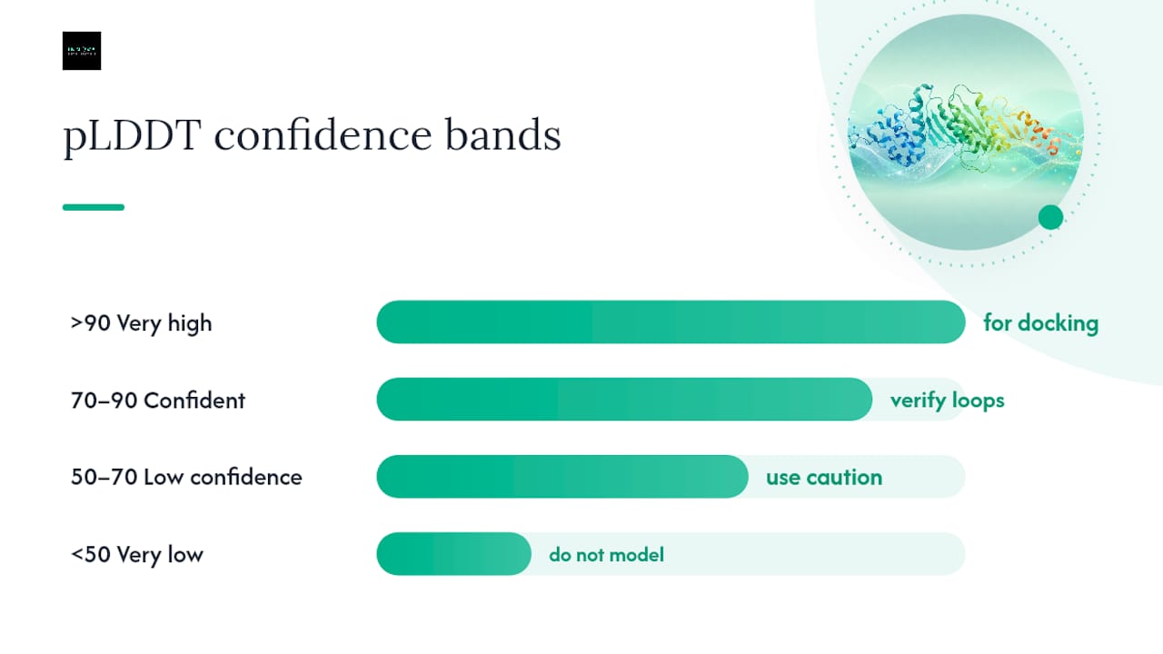
- pLDDT (per-residue local distance difference test): Ranges from 0–100. Scores above 90 indicate very high confidence; 70–90 are confident and generally reliable for most structural analyses; 50–70 are low confidence and should be treated cautiously; below 50 almost always flags an intrinsically disordered region rather than a modeling failure.
- PAE (predicted aligned error): A pairwise matrix reporting the expected positional error (in Å) between any two residues when the model is aligned on one of them. Low PAE between two domains means their relative orientation is reliable; high PAE means you should not trust inter-domain geometry for docking or design.
- pTM (predicted TM-score): Available in AlphaFold 3 and multimer outputs, it gives a global confidence score for the entire predicted fold or complex.
The quick rule: if your binding site sits in a region with pLDDT above 70 and low PAE to its neighboring secondary-structure elements, the model is a reasonable starting point. If the active site spans a low-pLDDT loop, plan for experimental validation before committing resources.
Key Takeaways
AlphaFold predicts 3D atomic protein models from sequence alone, but the pLDDT and PAE confidence scores determine whether a given prediction is ready for downstream research use or requires experimental validation first.
| Point | Details |
|---|---|
| What AlphaFold predicts | A 3D atomic model from amino-acid sequence, with per-residue pLDDT and pairwise PAE confidence scores. |
| Reading confidence correctly | pLDDT above 70 is generally reliable; below 50 signals likely disorder, not model failure. |
| Primary research uses | Hypothesis generation, functional annotation, docking prep, and mutagenesis design in high-confidence regions. |
| Core limitations | Low-MSA sequences, IDRs, membrane proteins, ligand binding, and conformational dynamics all reduce reliability. |
| Innovabiotech's role | Integrates AlphaFold predictions with MD refinement, virtual screening, and experimental design for drug discovery and protein engineering projects. |
Table of Contents
- Why AlphaFold changed structural biology overnight
- How does AlphaFold work, from sequence to structure?
- What AlphaFold outputs look like and how to read them
- How accurate is AlphaFold, and where does it fall short?
- How researchers use AlphaFold at proteome scale for functional annotation
- How to access AlphaFold predictions and the tools you need
- A checklist for using AlphaFold structures responsibly
- When and how to validate AlphaFold predictions experimentally
- AlphaFold in applied projects: what the confidence scores don't tell you
- Innovabiotech brings AlphaFold predictions into production-ready workflows
- Sources
- FAQ
Why AlphaFold changed structural biology overnight
The timeline is short and the impact is not. DeepMind's AlphaFold 2 won CASP14 in 2020 with accuracy competitive with experimental methods in many cases, a result that stunned a field that had spent decades on the protein-folding problem. The peer-reviewed Nature paper followed in 2021. Then, in 2022, EMBL-EBI and DeepMind released the AlphaFold Protein Structure Database with predictions for the human proteome and dozens of other organisms. The database now holds a very large number of predicted structures, each linked to UniProt entries and cross-referenced to the Protein Data Bank (PDB).
"AlphaFold has already been used to accelerate research into diseases such as malaria, Parkinson's, and antibiotic resistance — and the database is free and open to all." The scale of that resource, covering organisms from bacteria to humans, means that for many targets a researcher can retrieve a predicted structure within seconds rather than waiting months for crystallography.
The practical gains are real. Predicted structures accelerate hypothesis generation, enable large-scale functional annotation pipelines that would have been infeasible before the database existed, and give structural biologists a starting model for experimental phasing or cryo-EM fitting. EMBL-EBI curates AlphaFold DB, linking each entry to UniProt and related PDB content so predictions sit inside the broader biological context researchers already use.
How does AlphaFold work, from sequence to structure?
AlphaFold's prediction method is built on a few key ideas that separate it from classical homology modeling.
Inputs and evolutionary information
The model takes an amino-acid sequence and builds a multiple sequence alignment (MSA) by searching sequence databases for homologs. Correlated mutations in that MSA carry geometric information: if two residues co-evolve, they are likely in contact in the folded structure. AlphaFold also accepts optional structural templates from the PDB, though it can predict novel folds without close templates, which is what extends its reach into the "dark proteome" of proteins with no known structural relatives.

Architecture: Evoformer and the structure module (AlphaFold 2)
AlphaFold 2's core is the Evoformer, a stack of attention-based blocks that jointly processes the MSA and a pairwise representation of residue relationships. The Evoformer iteratively refines both representations, letting the model reason about physical and geometric constraints implicitly. A separate structure module then translates those representations into 3D backbone frames and side-chain torsion angles. The whole process runs through several recycling iterations, each pass refining the predicted coordinates.
AlphaFold 3: Pairformer and diffusion
AlphaFold 3 replaces part of the Evoformer with a Pairformer, reduces the weight given to MSA processing, and adds a generative diffusion module that outputs atomic coordinates directly. It also tokenizes ligands and modified residues, making it capable of modeling protein-ligand and protein-nucleic acid complexes. The trade-off: large complexes carry significant compute and memory costs.
Key inputs and outputs at a glance:
- Inputs: amino-acid sequence, MSA (from UniRef, BFD, and similar databases), optional PDB templates
- Processing: Evoformer/Pairformer attention layers, iterative recycling
- Outputs: 3D atomic coordinates (backbone + side chains), pLDDT per residue, PAE matrix, pTM for multimers
- Training data: Structures from the PDB; AlphaFold 3 also incorporates broader chemical data for ligand modeling
What AlphaFold outputs look like and how to read them
Every prediction from the AlphaFold DB or the AlphaFold Server comes with a standard set of files and metadata. Knowing what each contains saves time.
Output files you will encounter:
- PDB/mmCIF coordinate file: The 3D atomic model. The B-factor column in PDB format is repurposed to store pLDDT scores per residue, so any structure viewer that reads B-factors will color the model by confidence automatically.
- JSON metadata file: Contains the full pLDDT array, the PAE matrix, and (for AlphaFold 3 or multimer models) pTM scores. The PAE matrix is a 2D array of size N×N for an N-residue protein.
- Model ensembles: AlphaFold 3 and the AlphaFold Server can return multiple sampled structures, giving a rough sense of conformational spread.
pLDDT interpretation table:
| pLDDT range | Interpretation | Practical guidance |
|---|---|---|
| >90 | Very high confidence | Suitable for docking, mutagenesis design, and structural analysis |
| 70–90 | Confident | Generally reliable; verify loops at binding sites |
| 50–70 | Low confidence | Use with caution; cross-check with experimental data |
| <50 | Very low / likely disordered | Treat as intrinsically disordered; do not use for structural modeling |
Reading the PAE matrix
The PAE matrix is the most underused output in most workflows. A low PAE value between residue i and residue j (say, below 5 Å) means the model is confident about their relative position when the structure is aligned on either one. High off-diagonal PAE blocks between two domains mean the model has no reliable prediction for how those domains orient relative to each other, even if each domain individually scores well on pLDDT.
Pro Tip: Before running any protein-protein or domain-domain docking, open the PAE matrix. If the inter-domain PAE is high, treat each domain as an independent rigid body and dock them separately rather than using the full-chain model as a rigid template.
Downloading structures
The AlphaFold DB provides individual downloads by UniProt accession and bulk proteome packs (human, mouse, and many others) as gzipped tar archives. The AlphaFold Server accepts custom sequences and returns predictions within minutes for single chains. For programmatic access, the AlphaFold DB REST API returns JSON with coordinates and confidence data by accession.
How accurate is AlphaFold, and where does it fall short?
AlphaFold 2 achieved accuracy competitive with experimental methods in the CASP14 blind test, outperforming every other computational method by a substantial margin on median GDT_TS scores for single-domain proteins. For well-folded, single-chain proteins with deep MSAs, the predictions are often close enough to experimental structures that they can substitute for a starting model in molecular replacement or cryo-EM fitting.
That said, several categories of proteins and research questions expose real limits:
- Intrinsically disordered regions (IDRs): AlphaFold will produce coordinates for these, but low pLDDT is the signal that the region lacks a stable fold. The EMBL-EBI training guidance is explicit: low confidence often reflects genuine disorder, not model failure.
- Low-MSA sequences: Orphan proteins or highly divergent sequences with few homologs give the model less evolutionary information. Confidence scores drop, and the structural prediction becomes less reliable.
- Multimers and large assemblies: AlphaFold-Multimer handles homo- and heteromers, but large assemblies with many chains remain computationally expensive and less accurate than single-chain predictions.
- Ligand binding and post-translational modifications (PTMs): AlphaFold 2 predicts apo structures. It does not model bound ligands, cofactors, or PTMs, which can substantially alter conformation. AlphaFold 3 addresses this partially, but binding-induced conformational changes remain a challenge.
- Membrane proteins: Transmembrane regions can score well on pLDDT, but the lipid environment is absent from the prediction, and helix packing in the membrane context may differ from the predicted model.
- Conformational dynamics: AlphaFold predicts a single static model. For targets where multiple conformational states matter (kinases, GPCRs, allosteric enzymes), the static model captures one snapshot. MD refinement and binding-site analysis are necessary post-processing steps before using AlphaFold models in hit-to-lead optimization.
When to validate experimentally:
- Binding site spans a loop with pLDDT below 70
- Target is a membrane protein or large complex
- The research claim depends on precise side-chain geometry
- PTMs or cofactors are functionally critical
- The protein is known to be conformationally dynamic
Pro Tip: For drug-discovery targets, run a short MD simulation (50–100 ns) on the AlphaFold model before docking. This relaxes strained side chains, samples accessible binding-site conformations, and gives you a more realistic ensemble for virtual screening.
How researchers use AlphaFold at proteome scale for functional annotation
One of the less-discussed applications of AlphaFold is what it enables at scale: residue-level functional annotation across entire proteomes. Methods like PARSE use AlphaFold DB structures to compare predicted structures against known functional templates and propose candidate enzyme functions for uncharacterized proteins, including those in the "dark proteome" with no sequence-based annotation.
PARSE applied AlphaFoldDB to human and dark proteomes and generated numerous functional predictions after applying filtering rules to reduce spurious hits. The filters matter: without strict RMSD thresholds and residue-overlap requirements, low-complexity regions generate many false positives. The method reported that AlphaFold-predicted structures can improve downstream function-prediction models and in some benchmarks matched the performance of models trained on experimental structures for specific annotation tasks.
Practical caveats for this kind of pipeline:
- Low-complexity regions (coiled coils, repeats, IDRs) produce structural matches that are geometrically similar but functionally meaningless. Filter them out before experimental follow-up.
- Residue-level explainability matters: a structural match to an enzyme active site is only useful if the catalytic residues align, not just the overall fold.
- False-positive rates rise sharply when MSA depth is low. Apply an MSA-depth filter before including a prediction in a functional annotation pipeline.
These three filters together substantially reduce spurious hits before you commit to experimental follow-up. For genome-level annotation workflows, the functional annotation guide from Innovabiotech covers how these steps fit into pharma-grade pipelines.*
How to access AlphaFold predictions and the tools you need
Access options range from a browser search to a full local GPU installation, depending on your scale and compute resources.
- AlphaFold Protein Structure Database (alphafold.ebi.ac.uk): The fastest route for known proteins. Search by UniProt accession or gene name, download the mmCIF/PDB file and JSON confidence data, and you are working within a minute. Proteome packs for human, mouse, E. coli, and many other organisms are available as bulk downloads.
- AlphaFold Server (alphafoldserver.com): Submit a custom sequence and receive a prediction, including multiple sampled models and full confidence outputs. Useful for novel sequences not yet in the database. AlphaFold 3 is accessible here for protein-ligand and protein-nucleic acid complexes.
- ColabFold: A community-built Google Colab notebook that runs AlphaFold 2 (and related models) using MMseqs2 for fast MSA generation. It is the practical middle ground: no local GPU required, faster than the original pipeline, and free for moderate use. Researchers working with dozens of sequences at a time will find it more flexible than the server.
- Local AlphaFold installation: For large-scale runs (hundreds to thousands of sequences), a local installation on a GPU cluster is the only realistic option. The original DeepMind pipeline requires significant storage for sequence databases and a capable GPU (A100-class for reasonable throughput).
Visualization and analysis tools:
- PyMOL and UCSF ChimeraX both read mmCIF and PDB files natively and can color structures by B-factor (pLDDT) out of the box.
- Python scripts using Biopython or MDAnalysis can parse the JSON confidence files to extract pLDDT arrays and PAE matrices for programmatic analysis.
- For MD refinement post-processing, GROMACS and AMBER are the standard choices; both accept AlphaFold coordinate files with minor preparation steps.
When AI-assisted literature synthesis is part of your workflow for mining AlphaFold-linked discoveries, tools like PaperSynapse's AI literature synthesis guide can help you triage the rapidly growing body of AlphaFold-related publications efficiently.
A checklist for using AlphaFold structures responsibly
Before a predicted structure enters an experimental workflow or a publication, work through these steps in order:
- Inspect pLDDT per residue. Color the structure by pLDDT in PyMOL or ChimeraX. Identify any low-confidence regions (below 70) that overlap with your region of interest.
- Examine the PAE matrix. Download the JSON file and plot the PAE matrix. Confirm that the domain or region you plan to use has low PAE with its structural neighbors.
- Check MSA depth. If you ran the prediction yourself (ColabFold or local), check the number of effective sequences in the MSA. Fewer than 30 effective sequences is a warning sign for prediction reliability.
- Search for known experimental templates. Run a BLAST or structure-based search against the PDB. If a close experimental structure exists, compare it to the AlphaFold model before proceeding.
- Annotate predicted disordered regions. Use the pLDDT profile to flag IDRs explicitly in your working model. Do not include them in rigid-body docking or energy minimization without restraints.
- Ask the binding-site questions. Is the predicted active site or binding pocket in a high-confidence region? Are the key residues (catalytic, binding) in a low-PAE neighborhood? Is the region known to be dynamic?
- Cite correctly. Cite the AlphaFold Nature paper for the method and the AlphaFold DB for the database entry. Include the UniProt accession and the AlphaFold DB version in your methods section.
When and how to validate AlphaFold predictions experimentally
The type of experimental follow-up should match the specific claim you are making with the predicted structure.
- Fold verification: If the overall topology of a novel protein is the primary claim, X-ray crystallography or cryo-EM provides direct structural evidence. NMR is appropriate for smaller proteins (typically below 50 kDa) where solution-state dynamics also matter.
- Binding site validation: Biochemical binding assays (ITC, SPR, or fluorescence polarization) confirm that a ligand or partner binds where the model predicts. Site-directed mutagenesis of predicted contact residues tests whether those residues are functionally required.
- Functional assay: For enzyme function predictions from annotation pipelines, an activity assay with the proposed substrate is the most direct test. Structure alone does not confirm catalysis.
- Conformational dynamics: Hydrogen-deuterium exchange mass spectrometry (HDX-MS) maps solvent accessibility and flexibility across the sequence, directly testing whether predicted flexible regions behave as expected. MD simulations provide a computational complement and can sample conformational ensembles for hit-to-lead drug discovery before committing to synthesis.
Prioritizing which predictions to validate first:
- High pLDDT at the functional site + novel biology = highest priority for experimental follow-up.
- High pLDDT but known homolog with experimental structure = lower priority; use the experimental structure where possible.
- Low pLDDT at the functional site = validate the fold before any downstream work.
- Membrane protein or large complex = plan cryo-EM or native MS from the start; do not rely on the static model alone.
AlphaFold in applied projects: what the confidence scores don't tell you
The most common mistake in applied AlphaFold work is treating a high pLDDT score as a green light for everything downstream. It isn't. pLDDT measures local structural confidence, not functional relevance, not binding-competent conformation, and not the effect of the cellular environment on fold stability.
In practice, the most productive use of AlphaFold predictions is as a hypothesis-generation engine, not a replacement for experiment. A predicted structure with a well-defined binding pocket and high-confidence active-site residues tells you where to look and what to mutate. It does not tell you whether the protein is folded that way in the cell, whether a ligand will bind with useful affinity, or whether a PTM shifts the conformation enough to matter.
For teams running drug-discovery projects, the workflow that consistently works is: AlphaFold prediction, followed by MD refinement to sample binding-site conformations, followed by structure-based virtual screening, followed by targeted biochemical validation of the top hits. Skipping the MD step and docking directly to the raw AlphaFold model is a common shortcut that inflates false-positive rates in virtual screening. Innovabiotech's computational team integrates this full pipeline, including antibody design workflows and structure-guided protein engineering, for clients who need end-to-end support rather than just a predicted coordinate file.
Innovabiotech brings AlphaFold predictions into production-ready workflows
Retrieving an AlphaFold structure is the easy part. Turning it into a validated drug candidate, an engineered enzyme, or a designed peptide requires the computational and experimental integration that most research teams don't have in-house.

Innovabiotech's San Francisco-based team applies AlphaFold predictions directly in protein and chimeric protein engineering, enzyme optimization, and de novo peptide design projects, with MD refinement and structure-based virtual screening built into every drug-discovery engagement. Every project runs under a confidential workflow with full data security, and the team works project-by-project so you are not locked into a long-term retainer before you know the scope. To discuss how AlphaFold-guided structure analysis fits your specific target, reach out to Innovabiotech for a project scoping consultation.
Sources
- Highly accurate protein structure prediction with AlphaFold | Nature
- AlphaFold Protein Structure Database
- Enhancing Protein Function Prediction Performance by Utilizing AlphaFold-Predicted Protein Structures | PMC
- Enhancing Protein Function Prediction Performance by Utilizing AlphaFold-Predicted Protein Structures | Journal of Chemical Information and Modeling
FAQ
Why is AlphaFold a big deal for structural biology?
AlphaFold solved a 50-year-old problem: predicting protein 3D structure from sequence with accuracy competitive with experiment. The AlphaFold DB now provides over 200 million free predicted structures, giving researchers structural access to proteins that would take decades to solve experimentally.
Can AlphaFold predict protein structure from any sequence?
Yes, AlphaFold can generate a prediction for any amino-acid sequence, but confidence varies. Proteins with deep multiple sequence alignments and well-folded domains produce high-confidence models; intrinsically disordered proteins and low-MSA sequences produce low-pLDDT predictions that should not be used for structural modeling.
How does AlphaFold work, in simple terms?
AlphaFold searches for evolutionary relatives of your protein, extracts geometric information from their correlated mutations, and uses a deep neural network to translate that information into 3D atomic coordinates. It then reports how confident it is about each residue via the pLDDT score.
Is AlphaFold 100% accurate?
No. AlphaFold achieves near-experimental accuracy for many well-folded single-chain proteins, as demonstrated at CASP14, but it has documented limitations with disordered regions, membrane proteins, large complexes, and ligand-bound conformations. Always check pLDDT and PAE before using a prediction for experiments.
When should I validate an AlphaFold prediction experimentally?
Validate when the binding site or functional region has pLDDT below 70, when the target is a membrane protein or large multimer, when PTMs or cofactors are functionally critical, or when the research claim depends on precise side-chain geometry or conformational dynamics.