
Pharmacophore modeling maps the spatial arrangement of chemical features, hydrogen bond donors, acceptors, hydrophobic patches, and aromatic rings, that a molecule needs to bind a target, and drug discovery teams use it to identify hits, hop between chemical scaffolds, and filter docking results. The field has moved past static, single-conformation models: dynophores built from molecular dynamics and consensus approaches that pool multiple ligand or receptor states now define best practice for anything but the most rigid binding sites.
TL;DR:
- Ligand-based pharmacophore models are ideal when you have several diverse confirmed actives but lack structural data, emphasizing shared features across compounds.
- Structure-based models rely on crystal or cryo-EM structures, making them suitable when actual protein-ligand complexes are available, especially for novel targets.
- Hybrid approaches combine both data types to improve accuracy, especially when both ligand and structure information are present, but require cross-validation to prevent overfitting.
- Validating pharmacophore models demands using decoy sets and calculating metrics like enrichment factors and F0.5 scores to ensure they effectively distinguish actives from inactives.
- Incorporating molecular dynamics and AI-driven feature extraction enhances the modeling of flexible binding sites but increases computational costs, suitable for targets with known conformational variability.
Table of Contents
- What Is Pharmacophore Modeling and What Are the Main Types?
- How Do You Build a Pharmacophore Model Step by Step?
- What Are Dynophores and How Does AI Fit Into Pharmacophore Modeling?
- How Do You Validate a Pharmacophore Model?
- What Are the Most Common Pharmacophore Modeling Mistakes?
- Where Does Pharmacophore Modeling Actually Get Used?
- How Innovabiotech Approaches Pharmacophore Modeling Projects
- Should You Build a Pharmacophore Model or Bring in Outside Help?
- Get Expert Support for Your Pharmacophore Modeling Project
- Sources
- FAQ
What Is Pharmacophore Modeling and What Are the Main Types?
A pharmacophore model is not a molecule. It is an abstraction, a set of geometric constraints (a hydrogen-bond acceptor here, a hydrophobic sphere there, spaced at defined distances and angles) that describes what any molecule needs to have to bind a given target. That abstraction is what lets you screen a database of two million compounds in an afternoon instead of docking every single one.
Three approaches dominate practice, and picking the wrong one for your data situation wastes weeks.
Ligand-based pharmacophore modeling extracts shared features from a set of known active compounds, typically by aligning their bioactive conformations and identifying which features recur across the series. It works when you have no crystal structure but do have a reasonable set of confirmed actives, ideally 5 to 20 structurally diverse ones pulled from sources like ChEMBL or PubChem. The catch: the model only reflects what your training compounds happen to share, so a narrow or biased active set produces a narrow, biased pharmacophore.
Structure-based pharmacophore modeling derives features directly from a protein-ligand complex or, in receptor-based variants, from an apo (ligand-free) binding site. This sidesteps the training-set bias problem entirely because the features come from actual protein geometry rather than chemical inference. Tools like E-FTMap identify interaction hot spots by scattering small molecular probes across a binding site and finding energy minima, which is invaluable when no co-crystallized ligand exists yet.
Hybrid approaches combine both data streams, often anchoring ligand-derived features to a known binding pocket to correct for orientation ambiguity in the ligand-only model.
Choosing between them comes down to what data you actually have:
- Rich ligand SAR data, no structure: go ligand-based
- Crystal or cryo-EM structure, few or no known actives: go structure-based
- Both available: build hybrid, and cross-check one against the other before committing compute to screening
How Do You Build a Pharmacophore Model Step by Step?
The workflow is iterative by design. You rarely nail the final model on the first pass, and treating it as a one-shot exercise is how teams end up chasing false positives for months.
- Curate your data set. Pull actives from ChEMBL or PubChem, and generate matched decoys using a set like DUD-E so your validation set has property-matched, presumed-inactive compounds rather than random molecules that are trivially easy to distinguish.
- Enumerate conformers and stereochemistry. Generate a reasonable conformer ensemble per molecule, commonly in the range of 50 to 250 conformers within a 3 to 5 kcal/mol energy window above the global minimum. Too few conformers and you miss the bioactive pose; too many and alignment becomes computationally wasteful without added accuracy.
- Extract and align features. Identify chemical feature points across your aligned actives or your protein pocket, then cluster overlapping features into candidate pharmacophore points.
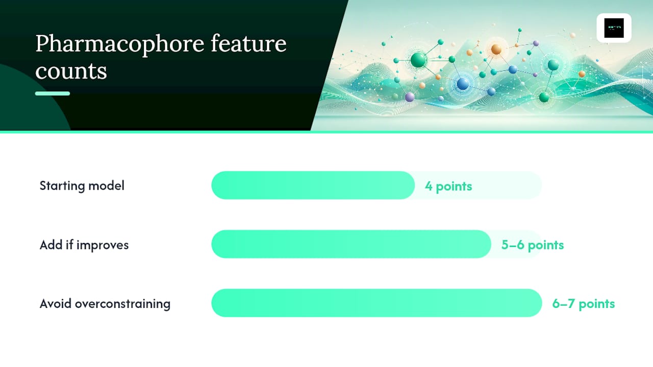
- Build the model incrementally. Start with a simple 4-point model (say, two H-bond acceptors and two hydrophobic centers) and only add a 5th or 6th point if it measurably improves enrichment without overfitting to a single active's conformation.
- Run the virtual screen. Apply a fast fingerprint-based prefilter to cut your library down, then run exact geometric matching against the survivors, and rank by fit score before triaging the top hits for visual inspection or docking.
Pro Tip: Resist the urge to add a 6th or 7th pharmacophore feature just because your software allows it. Every added constraint shrinks your hit list geometrically, and an overconstrained model built on one crystal pose will miss legitimate binders that engage the pocket slightly differently.
Your deliverable checklist should include the model file itself, a validation report with enrichment metrics, the exact decoy set used, and a full screening log documenting hit counts at each filtering stage. Without that paper trail, nobody, including future you, can reproduce the screen six months later.
What Are Dynophores and How Does AI Fit Into Pharmacophore Modeling?
Static pharmacophores assume a binding site holds still. Most don't. Loops flex, side chains rotate, and a pocket that looks closed in one crystal structure can open into a druggable groove a few nanoseconds later in a molecular dynamics trajectory. That gap is exactly what dynophores were built to close.
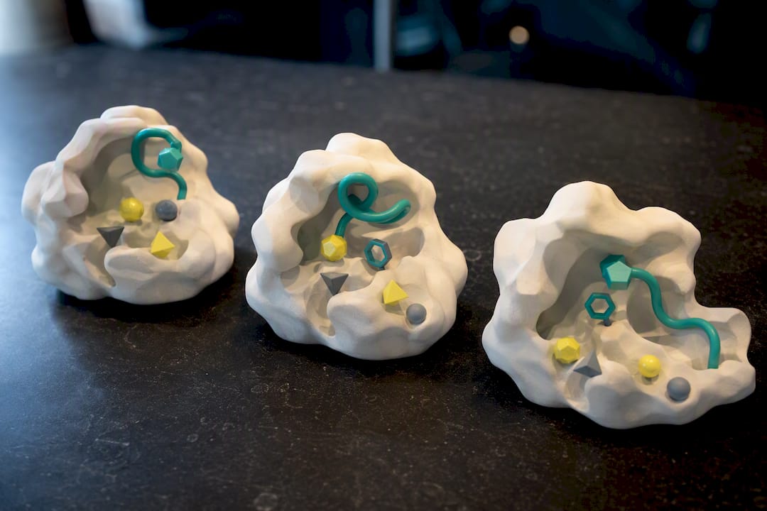
A dynophore captures how pharmacophoric features change across an MD trajectory rather than freezing them at a single frame, and recent field reviews frame this shift toward dynamic and consensus-based pharmacophore modeling as one of the defining advances in the discipline. Practitioner guidance is fairly direct on when this matters: the transition to MD-based ensemble models is often the deciding factor for success on flexible targets, where a single static structure would have led you astray.
Building one follows a predictable sequence:
- Run an MD simulation of the target, with or without a bound ligand, long enough to sample relevant conformational states
- Extract pharmacophoric features from evenly spaced snapshots across the trajectory
- Cluster those per-snapshot features by frequency and spatial consistency
- Retain only features that persist across a meaningful fraction of frames, discarding transient noise
- Assemble the surviving features into an ensemble or consensus pharmacophore
AI now plays a growing role in that middle step, automating feature detection and clustering across thousands of snapshots faster than manual inspection ever could, an approach with real parallels to how AI-assisted synthesis tools accelerate literature review by processing volume no human team can match manually. The caveat is the same one that applies to any automated pipeline: garbage in, garbage out. An ML feature extractor trained or tuned poorly will confidently flag noise as signal, so spot-check its output against a subset of manually curated frames before trusting it at scale.
None of this is free. A full MD-derived dynophore can cost days of GPU time versus minutes for a static ligand-based model, so reserve it for targets where flexibility is a known problem, allosteric sites, induced-fit pockets, intrinsically disordered regions, rather than defaulting to it everywhere.
How Do You Validate a Pharmacophore Model?
A pharmacophore model that hasn't been tested against decoys is a hypothesis, not a tool. Validation is where you find out whether your model actually discriminates binders from non-binders, or whether it just recovers the training compounds you built it from.
The standard approach uses a curated decoy set, DUD-E remains the most widely used, alongside a held-out set of confirmed actives never seen during model building. You run the screen, then score performance with a small set of standard metrics:
- F0.5 score, which weights precision over recall, is the right choice when you want a tight, high-confidence hit list; workflows commonly target F0.5 ≥ 0.8 for precision-focused selection
- F2 score, which weights recall over precision, suits early-stage screens where missing a true active is more costly than sifting a few extra false positives
- Enrichment factor (EF), reported at standard cutoffs like the top 1% or top 5% of the ranked library, to show how much better your model performs than random selection
Cross-validation across multiple training subsets, and consensus scoring across several independently built models, both reduce the risk that your validation numbers reflect a lucky split rather than genuine discriminatory power.
Whatever you report, report it reproducibly. A validation package worth publishing or handing to a collaborator includes the exact decoy set used, ROC curves, an EF table at multiple cutoffs, and the external test set composition, not just a single headline accuracy number that nobody can independently verify.
What Are the Most Common Pharmacophore Modeling Mistakes?
Most pharmacophore models that fail in practice fail for one of four predictable reasons, and each has a fairly direct fix.
Conformational and scaffold bias. A model built from actives that all share one chemotype will encode that chemotype's geometry, not the target's actual requirements. Expanding conformer sampling and building consensus models across chemically diverse active subsets, rather than one master model, reduces this single-hypothesis bias substantially.
Overfitting to geometric volume. A model with too many tightly constrained features will fit your training actives perfectly and reject everything else, including legitimate novel scaffolds. Adding shape-based exclusion volumes and testing rigorously against decoys catches this before it wastes a screening run.
Homogeneous training sets. If your actives all came from one chemical series or one publication, your model has learned that series, not the pocket. Cluster-based selection of training compounds, and pooling multiple independently derived models into an ensemble, corrects for this.
Data quality and compute limits. Public bioactivity databases contain assay noise, and MD-backed ensemble modeling is expensive. Prioritize where you spend compute: run full dynophore treatment on flexible or high-value targets, and reserve fast static pharmacophores for well-behaved, rigid pockets or early-stage triage.
Pro Tip: When a pharmacophore screen returns suspiciously few hits from a large library, don't assume the model is simply strict. Check whether one overly specific feature, often an aromatic ring constraint copied straight from a single crystal pose, is silently filtering out everything that doesn't match that exact orientation.
Where Does Pharmacophore Modeling Actually Get Used?
Three applications account for most of the field's real-world value, and each rewards a slightly different setup.
Virtual screening at scale is the most common use: a validated pharmacophore filters a library of millions down to a manageable shortlist in hours, feeding directly into the kind of large-scale screening pipelines that would be computationally impossible to run with docking alone on the full set.
Scaffold hopping plays to the pharmacophore's core strength: because the model describes an interaction pattern rather than a specific chemical skeleton, it can surface a completely different scaffold that satisfies the same geometric requirements, sidestepping a patent-blocked chemotype or a series with poor solubility.
Fragment-based expansion benefits from receptor-mapping methods like E-FTMap, which identify hot spots on the protein surface even without a bound ligand, then guide medicinal chemists on where to grow a fragment hit into a larger, higher-affinity molecule. This pairs naturally with structure-based fragment discovery workflows that use similar hot-spot logic.
For training and validation data, public repositories like ChEMBL, PubChem, and ZINC cover most early-stage needs. Most serious discovery programs eventually blend those public sets with proprietary in-house screening data once a project matures past initial hit identification, since internal SAR data reflects your actual chemical space far better than any public library.
How Innovabiotech Approaches Pharmacophore Modeling Projects
Every pharmacophore engagement at Innovabiotech starts with a scoping conversation, not a template. We define the inputs (target structure or active compound set, known SAR, prior screening data), the outputs (screening-ready model, ranked hit list, or both), the timeline, and the compute budget required, since a static ligand-based model and a full MD-backed dynophore campaign are different projects entirely.
Our approach to consensus and MD integration mirrors what's becoming standard across the field: build multiple independent pharmacophore hypotheses, cross-validate them against each other and against decoy sets, and only escalate to full molecular dynamics treatment when a target's flexibility genuinely warrants it. That keeps compute spend proportional to the problem rather than defaulting to the most expensive method on every target.
Deliverables include the model files themselves, a full validation report with enrichment and F-score data, decoy set documentation, and a screening log detailing every filtering stage, so a client's internal team can pick up the project and reproduce or extend it without starting from scratch.
Should You Build a Pharmacophore Model or Bring in Outside Help?
Pharmacophore modeling earns its place as your first computational step whenever you have a confirmed set of actives or a solved structure but haven't yet committed to a specific chemical series. It's fast, interpretable, and cheap relative to full docking campaigns or MD studies, which is exactly why it belongs early in a program rather than as an afterthought.
Layer in docking once you need binding-mode confidence on your top hits, and bring in dedicated MD or ML modeling when a target's flexibility is already a known problem. Most teams reach out for outsourced support at exactly that inflection point, when the target demands dynophore-level rigor but the internal compute or specialist bandwidth isn't there yet.
— Hooman
Get Expert Support for Your Pharmacophore Modeling Project
Building a validated, MD-backed pharmacophore model in-house means securing GPU time, a modeler who understands consensus clustering, and weeks you may not have before a program review. Innovabiotech runs these projects as a dedicated engagement, so you get a screening-ready model and a full validation package without diverting your own team from its current workload.

Our services cover ligand-based and structure-based pharmacophore generation, MD-backed dynophore construction for flexible targets, consensus modeling across multiple ligand or receptor states, and downstream hit-to-lead optimization once your screen returns candidates. A typical engagement includes a defined deliverable set (model files, enrichment and F-score validation reports, full screening logs), a project timeline agreed upfront, and confidential handling of all client compound and target data throughout. If your project also touches protein or enzyme structure work, we scope that alongside the pharmacophore work rather than as a separate contract. Reach out through Innovabiotech's virtual screening page to request a project estimate and timeline for your target.
Sources
For readers who want to go deeper on any single piece of this workflow, these sources cover the ground in more technical detail than a single article can:
- Pharmacophore modeling: advances and pitfalls
- Drug design by pharmacophore and virtual screening approach
- Pharmacophore modeling for targets with extensive ligand libraries (JOVE protocol)
FAQ
What Is an Example of a Pharmacophore?
A classic example is the pharmacophore for many kinase inhibitors: a hydrogen-bond acceptor and donor pair positioned to mimic ATP's interaction with the hinge region, combined with a hydrophobic feature occupying an adjacent pocket. The model doesn't specify a molecule, only that geometric arrangement, which is why chemically distinct kinase inhibitors can share the same underlying pharmacophore.
What Is the Difference Between Pharmacophore Modeling and Docking?
Pharmacophore modeling filters compounds based on whether they present the right features in the right geometric arrangement, while docking predicts a specific binding pose and estimates binding energy for each compound individually. Pharmacophore screening is typically faster and often used as a prefilter before docking the surviving candidates.
How Accurate Is Pharmacophore Modeling for Virtual Screening?
Accuracy depends heavily on model quality and validation rigor rather than the method itself; models validated with proper decoy sets and reporting enrichment factors and F-scores such as F0.5 ≥ 0.8 tend to perform far better than unvalidated models built on a narrow training set.
What Software Is Commonly Used for Pharmacophore Analysis?
Commercial platforms and open-source packages both support pharmacophore generation, feature extraction, and screening, with automated tools like PharmaCore-style pipelines increasingly used for structure-based model generation from co-crystallized ligand sets. Teams without in-house modeling infrastructure often outsource this work to specialized providers like Innovabiotech rather than building and maintaining the pipeline themselves.
Why Are Dynamic Pharmacophores (Dynophores) Becoming More Common?
Static pharmacophores miss binding-relevant conformational changes in flexible pockets, while dynophores built from molecular dynamics trajectories capture how interaction features shift over time. This makes them substantially more reliable for allosteric sites and induced-fit targets where a single crystal structure doesn't tell the whole story.